
- Molars
- Molars, located in the back of the mouth, are responsible for crushing and grinding food. Their complex anatomy features multiple roots, cusps, and a thick enamel coating. The first molars, which erupt around age 6, typically have three roots (two buccal and one lingual). The second molars, erupting around age 12, often have two or three roots, with the three-rooted form being more common. Third molars, or wisdom teeth, which erupt in late adolescence or early adulthood, can have a variable number of roots, ranging from one to four.
Anatomy of Teeth: Understanding the Building Blocks of Your Smile
Every time you flash a smile, you reveal a masterpiece of nature’s design: your teeth. These remarkable structures play a crucial role in our daily lives, from chewing food to articulating words. But what lies beneath the surface of our pearly whites?
Each tooth consists of three main parts:
Crown
The crown is the visible portion of the tooth that emerges above the gum line. It’s covered by enamel, the hardest substance in the human body, which protects the tooth from damage and decay.
Root
The root is the part of the tooth that anchors it to the jawbone. It’s embedded in the jaw and provides stability to the tooth. The root contains blood vessels and nerves that supply nourishment and sensation to the tooth.
Pulp
The pulp is the innermost layer of the tooth and contains blood vessels, nerves, and connective tissue. It’s responsible for the tooth’s vitality and growth. The pulp is protected by a layer of dentin, which is a hard tissue similar to enamel.
Understanding the anatomy of teeth is essential for maintaining good oral health. By knowing the different components of your teeth, you can better care for them and prevent problems like cavities and gum disease.
Types of Teeth: A Journey Through Your Pearly Whites
As we embark on the wondrous journey of oral health, let’s take a closer look at the cast of characters that reside in our mouths: the magnificent teeth. Each tooth, like a tiny masterpiece, serves a unique role in the symphony of our smile.
Central Incisors: The Charming Frontmen
At the very front of our dental stage, standing tall with their wide, rounded crowns, are the central incisors. These are the stars of our smile, responsible for presenting a welcoming beam.
Lateral Incisors: The Sidekicks
Stepping aside but equally charming are the lateral incisors, the sidekicks of the dental arch. They serve as the bridge between the central incisors and the more robust canines.
Canines: The Guardians
Next up are the canines, the fearless protectors of the smile. With their pointy, fang-like crowns, they’re designed to tear and shred food, safeguarding our precious smile from mishaps.
Premolars: The Hard-Working Duo
The premolars, also known as bicuspids, occupy the strategic position between the canines and molars. With two cusps or points on their crowns, they help break down food, preparing it for digestion.
Molars: The Crushing Powerhouses
At the back of the mouth, the towering figures of the molars take center stage. These robust teeth, with their broad, flat crowns studded with cusps, are the ultimate grinders, reducing food into a fine paste.
Molars: The Workhorses of Your Mouth
Molars, the mighty teeth nestled at the back of your mouth, are the unsung heroes of your dental game. Their robust anatomy empowers them to handle the toughest chewing challenges, grinding and crushing food into digestible bits.
Delving into the molars’ anatomy, we find a crown that proudly rises above the gum line, anchoring the tooth in place. The root, hidden beneath the gums, forms a solid foundation that keeps the molar firmly planted. Inside the tooth lies the pulp, a living tissue that houses nerves and blood vessels, nourishing the molar from within.
Molars don’t come in a one-size-fits-all mold. Our mouths house four types of molars: first molars, second molars, third molars (or wisdom teeth), and mandibular molars. Each type, with its unique shape and size, plays a specific role in the symphony of chewing.
First molars, emerging around age six, are the pioneers of the molar family. They possess a large chewing surface, adorned with four cusps, those pointy peaks that grip food and crush it with ease.
Second molars, their arrival marked at age 12, boast a similar anatomy to their first molar counterparts, but with a slightly smaller stature. They stand side-by-side with the first molars, forming a formidable crushing duo.
Third molars, better known as wisdom teeth, often make their grand entrance in the late teens or early twenties. These molars, shrouded in mystery, can vary in size and shape, sometimes adding an extra cusp to their armament.
Mandibular molars, residing in the lower jaw, are the perfect fit for their role. With narrower chewing surfaces and cusps adapted for a shearing action, they work harmoniously with the upper molars to grind food into a digestible paste.
The enamel covering molars is the strongest substance in the human body, a tough shield that protects the tooth from wear and tear. Beneath this protective layer lies a layer of dentin, a less brittle material that makes up the bulk of the tooth.
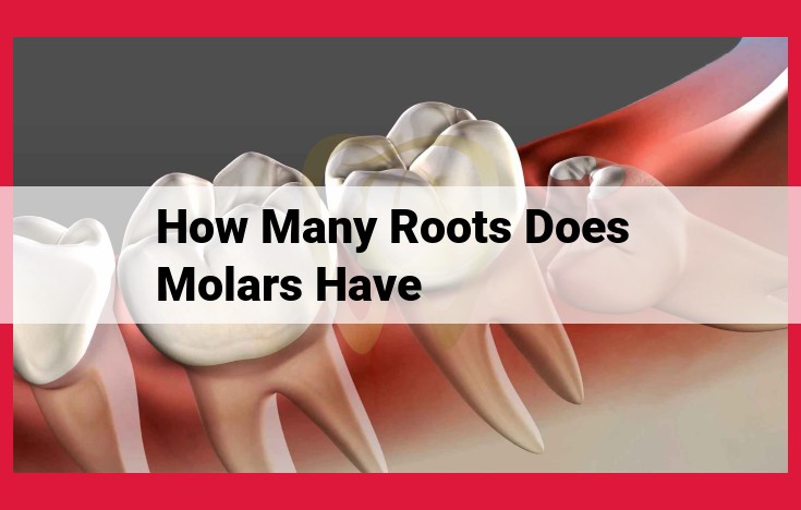
Root Morphology of Molars
The Anatomy of Molars:
Unveiling the intricacies of molars, we delve into their root morphology, a fascinating realm that holds significant implications in the realm of dental care. These powerhouses of mastication, strategically positioned in the posterior regions of our mouths, are characterized by their robust structure, designed to withstand the relentless forces of chewing.
Number of Roots:
Molars typically possess a generous number of roots, ranging from one to four. The most commonly encountered configuration is three roots, a testament to the molar’s exceptional stability. However, variations in root number do occur, with some molars exhibiting only two roots or even a solitary root.
Shape of Roots:
The shape of molar roots varies depending on the specific tooth. Maxillary molars (upper molars) typically have conical roots resembling slender cones. In contrast, mandibular molars (lower molars) often feature broader, flattened roots, providing a more substantial foundation.
Location of Roots:
The roots of molars are not haphazardly arranged but rather follow a precise pattern. The mesial root is located on the front side of the tooth, nearest to the midline of the mouth. The distal root lies on the back side of the tooth. Between these two primary roots, one or two additional accessory roots may be present, adding further support.
Importance of Root Morphology:
Understanding the root morphology of molars is paramount in the field of dentistry, as it directly influences treatment considerations and outcomes. For instance, the number of roots can impact the complexity and duration of root canal procedures. The **shape of the roots dictates the ease with which dental instruments can access the root canals. Moreover, the location of the roots guides the placement of dental restorations and implants.
Variations in Root Number
- Discuss the different variations in root number in molars, including the presence of extra roots or missing roots.
Variations in Root Number of Molars
delve into the captivating world of human teeth anatomy, exploring the intricate structures of the molar family. These formidable chewing machines are responsible for grinding and crushing our food, making their anatomical design a crucial aspect of our dental health. Like all aspects of the human body, molars exhibit a remarkable range of variations, including disparities in their root number.
Extra Roots: A Tale of Supernumerary Support
Among the most common variations is the presence of extra roots in molars. Typically, molars possess three roots; however, some individuals may have four or even five roots. These additional roots provide enhanced stability and support to the tooth, ensuring its ability to withstand the demanding forces of mastication. While extra roots are generally considered a positive trait, they can occasionally complicate dental procedures such as root canal therapy.
Missing Roots: A Puzzle of Asymmetry
In contrast to extra roots, some molars may have missing roots. This anomaly often affects the maxillary first molar, which may have two roots instead of three. Although less common than extra roots, missing roots can weaken the tooth, increasing its susceptibility to damage and decay. Missing roots may also need to be taken into consideration during dental treatments, such as orthodontic procedures or the placement of dental implants.
Clinical Significance: A Balancing Act
The number of roots in a molar has clinical implications that impact dental treatment decisions. For instance, molars with extra roots may require more time and expertise to extract, while those with missing roots may need additional support, such as crowns or bridges, to prevent breakage. Understanding the root number of a molar is essential for dentists to develop personalized treatment plans that ensure optimal oral health and longevity.
Research Frontiers: Unlocking the Mysteries
Research on root number variations in molars continues to shed light on the prevalence and impact of these anomalies. Studies have found that root number variations can vary significantly across different populations and geographical regions. Additionally, researchers are exploring the genetic and environmental factors that influence root number development, striving to unravel the mysteries surrounding these intriguing dental traits.
Clinical Significance of Root Number in Molars
The number of roots in molars plays a pivotal role in determining the complexity and prognosis of dental treatments. Understanding the clinical importance of root number is crucial for dentists in ensuring optimal patient care.
Endodontic Treatment
The presence of multiple roots can significantly complicate endodontic procedures. Each root contains a separate pulp canal, which must be identified, cleaned, and shaped during root canal treatment. The increased number of canals increases the risk of procedural errors, procedural accidents, such as perforation or stripping, and incomplete removal of infected tissue.
Surgical Interventions
In cases where tooth extraction becomes necessary, the number of roots influences the surgical approach. Molars with multiple roots require more extensive surgical intervention and may necessitate sectioning the tooth into smaller segments for atraumatic removal. This increases the risk of complications, such as damage to adjacent teeth or nerve structures.
Restorative Considerations
The root number also affects the prognosis of restorative treatments. Molars with fewer roots exhibit greater stability and resistance to occlusal forces. This makes them more suitable for large restorations, such as crowns or bridges. Conversely, molars with more roots have a higher risk of root fractures and may require additional support to ensure long-term success.
Prognostic Implications
The number of roots has implications for the overall prognosis of molars. Teeth with more roots generally have a better prognosis due to increased stability and resistance to forces. They are less likely to succumb to fractures or periodontal disease. In contrast, molars with fewer roots may be more susceptible to failure and may require closer monitoring and more frequent restorative interventions.
Research on Root Number of Molars: Unraveling Variations and Clinical Significance
Throughout our lives, our molars play a pivotal role in chewing and grinding food, making them essential for maintaining good oral health. While most of us have a basic understanding of tooth anatomy, the variations in molar root morphology can be fascinating and crucial for dental professionals to comprehend.
Prevalence of Variations
Extensive research has been conducted to understand the prevalence of variations in root number in molars. Studies have shown that up to 10% of molars can exhibit deviations from the typical number of roots. These variations can include extra roots (supernumerary roots) or missing roots (root fusion)
Impact on Treatment Outcomes
Variations in root number can significantly impact dental treatment outcomes. Molars with extra roots pose challenges in extraction procedures, requiring more complex techniques and potentially increasing the risk of complications. Conversely, molars with fused roots may be more susceptible to root canal infections due to the reduced number of access points for root canal treatment.
Recent Advancements
Advancements in dental imaging technologies, such as Cone-Beam Computed Tomography (CBCT), now enable dentists to visualize molar root morphology in unprecedented detail. This allows for more accurate diagnosis and tailored treatment plans, maximizing the chances of successful outcomes.
Understanding variations in root number is essential for dentists to provide optimal dental care for their patients. Research has shed light on the prevalence of these variations and their impact on treatment outcomes. By embracing advancements in imaging technology, dentists can effectively address variations in molar root morphology, ensuring the long-term health and functionality of our precious teeth.
